- Accueil
- Institut Néel
- Équipes de recherche
- Pôles & Services techniques
- Travailler à l’institut
- Partenariats
- Actualités
- Agenda
- Annuaire
The MRS (Materials, Radiation, Structure) team gathers researchers from complementary disciplines (physicists, chemists, geochemists, crystallographers), who develop and use cutting edge experimental and methodological tools, mastering the whole experimental process from synthesis, to physical and structural measurements. We share a marked specificity for extreme conditions studies and are strongly involved into CRG instruments at the Grenoble large facilities (ESRF, ILL). The systems under study range from magnetoelectric oxides to intermetallic materials for energy, through semiconductor nanowires, cultural heritage materials and hydrothermal fluids.
 |
 |
 |
See also the 2025 MRS presentation Poster
We use a large number of synthesis methods to prepare materials in the form of nanoparticles, polycrystalline powders or single crystals: controlled atmospheres, high pressure/high temperature, soft chemistry, topochemistry, hydrothermal conditions, flow or vapour transport.
The variety and development of these methods are necessary to obtain a large number of metastable phases (kinetically but not thermodynamically stable).
 |
 |
> See also « Discovery of a novel superconducting iron-based silicide: LaFeSiO1-δ« and the corresponding poster
We are studying semiconductor nanowires by TEM, in order to understand their properties, and in particular to couple them at the level of a single object.
In thin films, magnetic and electronic phases are stabilised by the epitaxial stresses of the substrate. We are interested in linking these new properties measured macroscopically (resistivity) to their stress structures seen by Raman scattering and X-ray diffraction with a view to fine stress engineering.
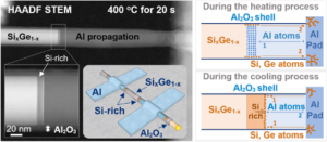
Example of an in-situ experiment where a solid state reaction between an Aluminum contact on a SiGe nanowire is started using
Joule heating and studied in-situ in the TEM. (Link to publication: 10.1021/acsanm.0c02303).
See also « Reversible diffusion of aluminium and silicon atoms in a nanowire » and the corresponding poster
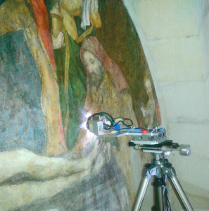
Our studies of heritage materials enable us to acquire new knowledge by identifying the materials used and also to describe their degradation over large time scales. These analyses contribute to the rediscovery of technical know-how and the use of certain raw materials, leading to the development of new ‘archaeomimetic’ materials.
See also « Revealing the techniques of medieval artists by X-ray analyses » and the corresponding poster
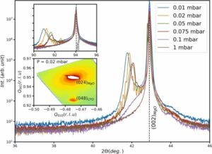
The study of the temperature and pressure dependence of the structure and collective dynamics of glass-forming materials at the atomic scale uses coherent X-ray scattering (XPCS) techniques at ESRF under high pressure.
See also « Glasses by X-ray Photon Correlation Spectroscopy » and the corresponding poster
Heterogeneous materials contain different materials with dramatically different mechanical and physical properties. They can be defined as materials with dramatic heterogeneity in strength from one microstructural domain to another. This strength heterogeneity can be caused by microstructure heterogeneity, crystal structure heterogeneity, or compositional heterogeneity. The domain sizes could range from micrometers to millimeters, and the domain geometry can vary to form very diverse material systems. Many natural and synthetic heterogeneous materials exist, such as bone, wood, animal tissues, plant cells, sands, soils, multiphase composites, concretes, sandwich structures, foams, and multi-layered structures. These materials are widely utilized as both structural and functional components in different devices. Until recently, scientists’ general procedure to understand the chemistry and physics of heterogeneous systems was to study large and complex structures and then examine the fundamental and smaller building blocks of those structures. This approach is called top-down science. However, with the development of scanning probe microscopes allowing the observation of individual atoms and molecules, it became possible to design and build new structures from their atomic-level constituents, one atom or one molecule at a time, i.e., “Materials according to the design”. This ability to carefully arrange atoms offers new opportunities to develop mechanical, electrical, magnetic, and other properties that would otherwise be impossible. We call this the bottom-up approach, and the study of the properties of these materials is called nanotechnology.
See also « Towards better industrial catalysts for reducing air pollution » and the corresponding poster
Hydrothermal fluids circulating in the Earth’s crust are essential for the transport of metals and their deposition in the form of deposits. On the FAME light line we have developed methods based on X-ray absorption spectroscopy to determine the molecular structure and concentration of the metal complexes responsible for this transport.
Raman measurements are also carried out to characterise solvent structures under hydrothermal conditions. All these in situ experiments are based on the development of dedicated autoclaves.

We are developing measurement and analysis techniques to give us an original perspective on our themes.
At the synchrotron, we are developing :
Using transmission electron microscopy, we are developing 3D diffraction methods to determine crystalline structures, and characterising electric fields using electron holography, 4D STEM and ptychography, with in situ biasing.
As part of the interdisciplinary CDP PATRIMALP project, we are developing original instruments such as Mobidiff for non-invasive fluorescence and X-ray diffraction measurements.
For all these techniques, we are adapting analytical strategies and innovative methods for processing massive imaging data using Artificial Intelligence, adapted to the analysis of nano-objects or ancient and heritage materials.
The different methodological developments at the MRS team:
The understanding of the physical properties of a material often requires a thorough knowledge of its structure. However, there is a large number of materials for which the ab initio determination of the crystallographic structure is not possible using powder or single crystal X-ray diffraction, due to the complexity of the powder diffraction pattern, the nanostructured aspect of the materials (nanoparticles, nanowires, thin films, …) or the difficulty to synthesize single crystals with a sufficient size.
During the last decades, the impressive progress of the technical performances of transmission electron microscopes has led to significant advances in physics, material science and life sciences. Thanks to the fabulous progresses in Electron Crystallography since the invention of electron beam precession in 2008 and its use in Precession Electron Diffraction Tomography (PEDT) new methods based on electron diffraction allow to solve ab initio crystal structures by calculating a first structural model and refining it with a dynamical refinement. These techniques are nowadays complementary to X-ray diffraction and/or neutron diffraction and perform on a comparable level. The huge advantage of PEDT is to exploit the specificity of electron diffraction, perfectly adapted to the study of single crystals of a few tens of nanometers in diameter, i.e. whose volume is 106 times smaller than the one necessary for X-ray diffraction on single crystals. In addition, the use of electron diffraction is more favorable than X-ray diffraction for materials with low irradiation resistance, because it is sufficient to apply an amount of irradiation 103 to 104 times smaller than in X-ray diffraction to obtain the same useful signal.
The combination of these two advantages places electron crystallography at the forefront of techniques suitable for structure determination of many materials, especially beam sensitive materials such as MOFs, zeolites, organic compounds, light elements battery materials.
Low Dose Electron Diffraction Tomography (LD-EDT)
In this context, we have developed an innovative method of low dose electron diffraction tomography (LD-EDT) which requires only a very low irradiation dose (less than 1 e-/Ų) [1] to obtain a data set leading to the structure of a crystal. The electron dose received by the sample is minimized by working with a very weak incident beam and by discarding the beam from the sample during all times other than the recording of the diffraction intensities.
In this method, the crystal is tilted in steps around the goniometer axis and a diffraction pattern representing a slice of the reciprocal space is recorded at each step position. From these slices the 3D reciprocal space is reconstructed and the diffracted intensities are measured.
From the data obtained and using specialized crystallographic software, the structures of different crystals were solved in cases where X-ray diffraction didn’t succeed. Refinement taking into account the dynamical theory of diffraction can be performed on the data and reveals the fine details of the crystals.
This method is particularly efficient for metal-organic frameworks (MOF), zeolites or organic crystals, which are too complex to allow the determination of their atomic structures from X-ray powder diffraction data (large unit cell, low symmetry, large number of atomic positions).
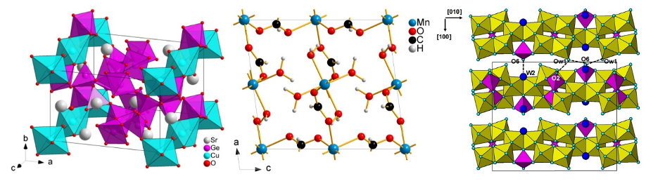
Figure 1: examples of structures solved by LD-EDT
Continuous 3D Electron Diffraction
Another method to prevent the material deterioration under the electron beam during the data collection is to work faster by recording a video in ED mode during the continuous sample holder tilt. The experiment is performed using a single tilt tomography sample holder with a tilt range of -55°/+53°. Once the movie recorded, the individual ED patterns are then extracted from the camera software options, and are processed by the conventional crystallography software consisting in peak identification, 3D reciprocal space reconstruction, intensities extraction and structure calculation. Thanks to this method, the crystal structure of the potential battery material Na2VO(HPO4)2 has been solved (Figure 2) [2].
See also « Combining electrons and X-rays to solve the structure of a new battery material » and the corresponding poster
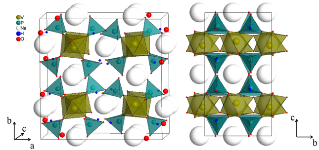
Figure 2 : Na2VO(HPO4)2 crystal structure
[1] S. Kodjikian and H. Klein, 2019, Ultramicroscopy, 200, 12-19, https://doi.org/10.1016/j.ultramic.2019.02.010
[2] C. Lepoittevin, O. Leynaud, A. Neveu, T. Barbier, M. Gnanavel, V. Gopal, V. Pralong, Dalton Trans., 2021, 50, 9725–9734.
Fourier ptychography is a variation of the idea of the synthetic aperture microscopy (SAM) method. Instead of scanning the sample, it is illuminated from different angles, and a bright-field image is recorded at each angle. These images are therefore combined to extend the numerical aperture of the microscope objective and thus improve spatial resolution without resorting to fluorescent markers. Fourier ptychography is therefore a super-resolution method which exceeds the diffraction limit and which has the advantage of being a non-invasive method, without marking and with a very wide field of view.
In view of the principle of reciprocity of microscopy, this method is the Fourier counterpart of conventional ptychography, where the locations of the sample plane and the plane of the aperture are effectively switched. This is a bright field imaging approach that uses images generated from illumination at different angles, with overlap in Fourier space of the contrast transfer function to the retro-focal plane of the lens, to create an image with enhanced SBP. This overlap introduces redundancy in the data, which makes it possible to simultaneously obtain images of intensity and phase contrast of the sample, as well as the pupil function of the microscope through an iterative algorithm, derived from that of X-ray ptychography.
To achieve 3D X-ray imaging, the method typically used is Computed Tomography (CT). Most of the X-ray imaging technique will enable 2D imaging, but in combination with CT, one can obtain 3D images of the sample. The 2D imaging experiment is repeated for each tomographic angle, by rotation the sample typically, over 180 or 360 degrees. By cast the 2D images into tomographic reconstruction algorithms, the volumetric imaging of the sample is obtained. For this reason and as examples, we will call the techniques as follows: Diffraction tomography (diff-tomo), Phase Contrast Imaging (PCI) tomography, holographic tomography, speckle-tracking tomography, Coherent Diffraction Imaging (CDI) tomography, and Ptychographic X-ray Computed Tomography (PXCT).
X-ray Ptychography (also referred as far-field X-ray ptychography) is a coherent diffractive imaging technique capable of providing highly detailed images of a sample’s complex-valued transmittance. A collection of coherent diffraction patterns is generated by scanning across the sample with a spatially confined illumination and sufficient overlap of adjacent illumination footprints. These overlapping illuminations introduce redundancy in the data, which is exploited to simultaneously provide information on specimen and illumination. The spatial resolution is not limited by the incoming beam size, but rather by the highest scattering vector in which speckles can be detected. The ptychographic phase retrieval consists in an iterative algorithm which will reinforce consistency between each scan position and the corresponding diffraction pattern, linked by Fourier transforms, as well as consistency between the adjacent scan positions due to the overlap. This redundancy allows us to simultaneously retrieve the amplitude and phase shifts of the wavefield past the object and the incoming wavefield function (probe).
Near-field X-ray ptychography (NFP) is a variant of far-field X-ray ptychography in the holographic regime which has recently been implemented at the ID16A beamline of the ESRF. NFP relies however on a magnification geometry, in which the sample is positioned in a defocused position downstream of focus of a divergent beam to achieve a given pixel size. Consequently, the spatial resolution will be limited by the focus size upstream the sample, different from the far-field ptychography. However, if the beam focus can be small as in the ID16A beamline, near-field ptychography allows us to measure a larger field-of-view than the far-field version in less time which is a big advantage for the nanoimaging of large scaled-up materials. NFP is able to retrieve a sample’s complex-valued transmission function from multiple near-field diffraction images, each with a lateral shift of the sample within the 1st Fresnel Zone and with a structured illumination. The structured illumination brings transverse diversity: i.e., the scattering direction changes at different lateral positions in comparison to the scattering of the object in plane waves. The phase retrieval in NFP is very similar to that of far-field X-ray ptychography, with the difference that the consistency between each scan position and the diffraction pattern is linked by Fresnel transforms rather than by Fourier transforms.
 |
The coupling between spin-charge-lattice-orbital degrees of freedom is at the origin of intriguing phenomena in strongly correlated systems. For instance, in frustrated magnets, both the geometry of the lattice and the nature of the magnetic exchange interactions are source of interesting physical phenomena such as non-trivial order, metamagnetism, spin liquid, etc … |
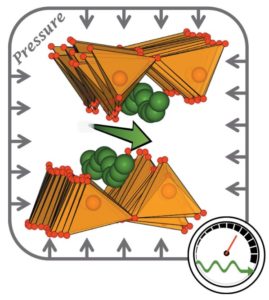
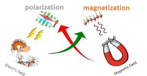
Our research is focused on a class of solid materials known as « multiferroics, » which exhibit coexistence of magnetic and electrical orders that are intertwined through the co-called magnetoelectric coupling. Such effect enables the control of polarization (magnetization) by the application of a magnetic field (electric field) and makes multiferroics very attractive for potential applications.
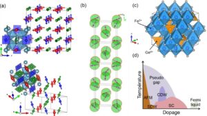
Another means of realizing unusual emergent properties is by tuning a phase transition, such as magnetic order, by modifying a non-temperature parameter: pressure, magnetic field, or chemical composition. Under these conditions, critical fluctuations result in unusual states such as unconventional superconductivity or exotic magnetic arrangements.
Our target materials include cation-ordered perovskites, spinels, pyroxenes, layered systels, and mixed anion compounds. In order to elucidate the underlying microscopic mechanisms that underpin the emergence of all these properties, we adopt an integrated approach, encompassing the design of new phases, their synthesis, and the advanced characterisation of their structural and magnetoelectrical properties. Macroscopic characterisations conducted in the laboratory are combined with state-of-the-art techniques on large-scale facilities, including elastic and inelastic neutron scattering and X-ray spectroscopy.
See also « Low dimensionality and frustration promote multiferroicity in SrMnGe2O6 pyroxene single crystal » and the corresponding poster
The entire field of metal hydrides for hydrogen storage and conversion is covered, including the search for new metastable phases, the functionalisation of materials through specific microstructures, and the integration of these materials into optimised heat exchange systems. In the search for new hydride compounds, we are studying the pressure-temperature phase diagram of ABH3 perovskite, high-entropy alloys and hydrogen absorption activation processes, storage processes and control systems. Our current research activities :
|
|
 |
|
|
Operando investigation of Electrochemical Flash Sintering of a self-supported All-Solid-State Battery monolith by total scattering (with a 50 ms Bragg and PDF temporal resolution) |
Permanents: Aude Bailly, Maria Diaz Lopez, Laetitia Laversenne, Patricia de Rango
Students, Post-doc, and Visitors: Léa Abou-Samra, Vishnu Dhinakaran, Riku Fukada, Zéphyr Glangeaud Massa, Arnaud Griffond, Gabriel-Ricardo Gomez-Eslava, Antonin Raynal
Strong interactions with the following technological pools: Cryogénie, Ingénierie Expérimentale, Automatisation et Caractérisation, X-Press
The different characterization methods at the MRS team:
We use several x-ray techniques in resonance condition, when the energy of the photons is tuned to an inner energy level of an atom. A substantial increase in scattering can be observed. The resonance consists in the absorption of a photon by a core electron promoted to one unoccupied orbitals sensitive to the local structural, electronic and magnetic structure. If this photoelectron then comes back to its core state, with no energy left behind, the process is said to be “resonant elastic x-ray scattering” (REXS) and can give rise to diffraction. The resonance can also trigger collective or individual excitation among the nearby electrons and the system can instead be left with some excitations. The emitted photon has then less energy, that’s “resonant inelastic x-ray scattering” (RIXS), which gives invaluable informations about the electronic or magnetic interactions. REXS or RIXS are photon-in photon-out scattering techniques, and thus can allow very versatile sample environments, like cryostats, oven, electric and magnetic field applications. Synchrotron facilities are required for tuning the beam to the desired energy. There, the very high flux of photon allows microscopic crystal to be measured, as well as ultrathin films.
Resonant X-ray Diffraction (RXD) is a REXS technique that consists in following the intensity of the diffraction of a crystal when the energy is swept through a resonance. RXD combines x-ray diffraction and x-ray spectroscopy. With this sensitivity, the resonant reflections carry information on the long-range electronic and magnetic states. New diffraction conditions can reveal superstructures invisible by other techniques revealing hidden orders in crystalline materials at the origin of macroscopic properties, electric or magnetic. RXD unraveled and quantified charge ordering in magnetite, and orbital and magnetic ordering in manganites. We perform RXD at D2AM at the ESRF, SEXTANTS at Soleil, I16 at Diamond. We use FDMNES and Dyna for the analysis of our data.
Contact: Stéphane Grenier (contact here)
Resonant Inelastic X-ray Scattering (RIXS) consists in measuring the difference in energy and the change in direction of the emitted photon from the incident photon. The difference in energy corresponds to the energy used by all sort of excitations, charge, magnetic like magnons or for collective vibrations, like phonons. The change in direction indicates the propagation of the excitations relatively to its crystal lattice axes. Mapping the excitations offers a view on the interactions at play in the system. We used RIXS to measure magnons and charge-phonon couplings in high-Tc superconductors and crystal field excitations in multiferroics. We performed RIXS at ID32 at the ESRF and SEXTANTS at Soleil.
Contact: Laura Chaix (contact here)
Y. Joly, S. D. Matteo, and O. Bunău, Resonant X-Ray Diffraction: Basic Theoretical Principles, Eur. Phys. J. Spec. Top. 208, 21 (2012).
J. Fink, E. Schierle, E. Weschke, and J. Geck, Resonant Elastic Soft X-Ray Scattering, Rep. Prog. Phys. 76, 056502 (2013).
J. E. Lorenzo, Y. Joly, D. Mannix, and S. Grenier, Charge Order as Seen by Resonant (Elastic) X-Ray Scattering, Eur. Phys. J. Special Topics 208, 121–127 (2012).
L. J. P. Ament, M. van Veenendaal, T. P. Devereaux, J. P. Hill, and J. van den Brink, Resonant Inelastic X-Ray Scattering Studies of Elementary Excitations, Rev. Mod. Phys. 83, 705 (2011).
X-ray Resonant Magnetic Scattering, Dyna project.
Dyna is a simulation and fitting program for x-ray and optical reflectivity and transmittance. Its specificity lies in the account of anomalous, resonant and magnetic resonant effects and it is used for the study of structural, magnetic and electronic stackings in ultrathin multilayers.
Dyna is a collaborative, open and free program, with collaborations at Institut Néel, Soleil synchrotron, and Sorbonne University. Visit the webpage of the projet dyna for documentation, and give it a try !
Contact: Stéphane Grenier (contact here)
The MRS group is engaged in the elaboration of a large variety of materials including compounds with unfavorable thermodynamic stability (metastable phases or with uncommon oxidation states), that range from multi-element oxides, chalcogenides, iron-based pnictides, intermetallics and hydrides, molecular framework solids or even composites. This activity benefits from a large panel of techniques available on-site for the design of single-crystals, ceramics or nano-materials in collaboration with three technical groups at Néel (“X’Press”, “Cristaux massifs” and “ThEMA”).
solid-state reactions routes with glove-box for manipulation in water/O2-free conditions combined to thermal treatments in inert or reactive atmosphere: from vacuum to controlled gas mixtures and 1600°C maximum temperature
solution-based techniques under milder conditions including coprecipitation, polymeric and/or auto-combustion routes; colloidal chemistry
crystal growth by chemical vapor transport
vacuum induction melting for reactive metals and alloys
pressure-assisted synthesis with fondant or oxidizing agent, using two large volume presses reaching 7 GPa and 1200°C; possibility for a preliminary screening of the suitable (T,P) conditions from in-situ powder x-ray diffraction combined to Paris-Edinburgh press or in-house designed pressure cell with independent control of temperature and pressure
high-energy mechanical milling and melt spinning
hydrogenation post-treatment through solid-gas reaction, possibly combined to severe plastic deformation processes to change sorption properties
We use several TEM based techniques to learn more about materials, and in parallel also develop these techniques to make them more quantitative. The focus lies on crystallography of particles that can’t be studied by X-rays (due to size or other difficulties) by 3D diffraction techniques, as well as studying electrical properties at nm and atomic length scales, for which we may want to electrically contact the sample. For these studies, we focus on 4D scanning TEM and analyze the diffraction maps both with more conventional methods (Center of Mass or template matching) as well as electron ptychography. Moreover, additional insight may be gained from off axis electron holography. We have developed a TEM compatible silicon nitride membrane technology with the Nanofab cleanroom allowing us to contact nano objects or thin flakes using lithography techniques for in-situ biasing and heating (S)TEM studies.
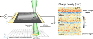 See also « Imaging electricic fields at nanometer length scales »
Contacts :
Crystallography : Holger Klein (holger.klein@neel.cnrs.fr) & Christophe Lepoittevin (christophe.lepoittevin@neel.cnrs.fr)
Field mapping and in-situ : Martien den Hertog (martien.den-hertog@neel.cnrs.fr) https://neel.cnrs.fr/les-chercheurs-et-techniciens/martien-den-hertoghome
Ptychography : Julio Cesar DaSilva (julio-cesar.da-silva@neel.cnrs.fr) https://neel.cnrs.fr/les-chercheurs-et-techniciens/jdasilva
Publications :
B.C. da Silva, Z.S. Momtaz, E. Monroy, H. Okuno, J.L Rouvière, D. Cooper and M.I. den Hertog. Assessment of Active Dopants and p–n Junction Abruptness Using In Situ Biased 4D-STEM, Nano Letters 22,23, 9544–9550 (2022) https://dx.doi.org/10.1021/acs.nanolett.2c03684
B.C. da Silva, Z.S. Momtaz, L. Bruas, J.L Rouvière, H. Okuno, D. Cooper and M.I. den Hertog. The influence of illumination conditions in the measurement of built-in electric field at p–n junctions by 4D-STEM, Applied Physics Letters 121, 123503 (2022) https://doi.org/10.1063/5.0104861
M.I. den Hertog, F. Donatini, R. McLeod, E. Monroy, C. Sartel, V. Sallet and J. Pernot. In-situ Biasing and Off-axis Electron Holography of a ZnO Nanowire, Nanotechnology 29 025710 (2018) https://doi.org/10.1088/1361-6528/aa923c
Many researchers of our team are involved in the scientific activity of synchrotron and neutron national equipments (Collaborative research groups) located in the European ESRF and ILL facilities. The three instruments that concern our group are :
![]() D2AM at ESRF, dedicated to structural investigations using anomalous scattering in materials science, with two instruments : a Kappa diffractometer and a small angle scattering camera.
D2AM at ESRF, dedicated to structural investigations using anomalous scattering in materials science, with two instruments : a Kappa diffractometer and a small angle scattering camera.
![]() FAME at ESRF, dedicated to x-ray absorption experiments for structural investigation of (very) diluted systems of environmental, material and biological interest.
FAME at ESRF, dedicated to x-ray absorption experiments for structural investigation of (very) diluted systems of environmental, material and biological interest.
![]() D1B at ILL, which is a high intensity powder diffractometer designed for in situ experiments and magnetism studies.
D1B at ILL, which is a high intensity powder diffractometer designed for in situ experiments and magnetism studies.
Position type: Stages Master-2 & Thèse
Contact: TOULEMONDE Pierre - 0476887421
Layered Lnn+1NinO3n+1 nickelates, for some of them, become high Tc superconductors under high pressure, after a structural transition occured. In this internship we will focus our study on n=1 stoichiometric La2NiO4 and n=2 substituted (La2Ln)Ni2O7 (with Ln=Sm,Pr) oxides. The aim is to understand the close relationship between superconductivity and the crystallographic transition, then the related superconductivity mechanism. Another goal will be to stabilize this superconducting state at ambient pressure in such nickelates family.
Person in charge: Martien Den Hertog, Stéphane Grenier
Permanents
Students & Post-docs & CDD
Aude BAILLY
Personnel Chercheur - CNRS
aude.bailly [at] neel.cnrs.fr
Phone: 04 76 88 90 19
Office: F-412
Pierre BORDET
Personnel Chercheur - CNRS
Pierre.Bordet [at] neel.cnrs.fr
Phone: 04 76 88 74 24
Office: F-407
Nassira BOUDJADA
Personnel Chercheur - UGA
Nassira.Boudjada [at] neel.cnrs.fr
Phone: 04 76 88 74 11
Office: F-207
Pierre BOUVIER
Personnel Chercheur - CNRS
pierre.bouvier [at] neel.cnrs.fr
Phone: 04 76 88 79 90
Office: F-402
Laura CHAIX
Personnel Chercheur - CNRS
laura.chaix [at] neel.cnrs.fr
Phone: 04 76 88 11 42
Office: F-413
Claire COLIN
Personnel Chercheur - UGA
claire.colin [at] neel.cnrs.fr
Phone: 04 76 88 74 14
Office: F-410
Julio Cesar DA SILVA
Personnel Chercheur - CNRS
julio-cesar.da-silva [at] neel.cnrs.fr
Phone: 04 76 88 11 06
Office: F-210
Céline DARIE
Personnel Chercheur - G-INP
Celine.Darie [at] neel.cnrs.fr
Phone: 04 76 88 79 40
Office: F-310
Martien DEN-HERTOG
Personnel Chercheur - CNRS
martien.den-hertog [at] neel.cnrs.fr
Phone: 04 76 88 10 45
Office: F-313
Fabio DENIS-ROMERO
Personnel Chercheur - CNRS
fabio.denis-romero [at] neel.cnrs.fr
Phone: 04 76 88 78 05
Office: F-309
Patricia DERANGO
Personnel Chercheur - CNRS
patricia.derango [at] neel.cnrs.fr
Phone: 04 76 88 90 30
Office: V-115
Maria DIAZ-LOPEZ
Personnel Chercheur - CNRS
maria.diaz-lopez [at] neel.cnrs.fr
Phone: 04 76 88 11 42
Office: F-413
Eric DOORYHEE
Personnel Chercheur - CNRS
Eric.Dooryhee [at] neel.cnrs.fr
Phone: 04 76 88 90 10
Office: F-417
Stéphane GRENIER
Personnel Chercheur - CNRS
stephane.grenier [at] neel.cnrs.fr
Phone: 04 76 88 90 98
Office: F-412
Jean-Louis HAZEMANN
Personnel Chercheur - CNRS
Jean-Louis.Hazemann [at] neel.cnrs.fr
Phone: 04 76 88 74 07
Office: F-419
Jean-Louis HODEAU
Personnel Chercheur - CNRS
Jean-Louis.Hodeau [at] neel.cnrs.fr
Phone: 04 76 88 11 42
Office: F-413
Olivier ISNARD
Personnel Chercheur - UGA
Olivier.Isnard [at] neel.cnrs.fr
Phone: 04 76 88 11 46
Office: F-203A
Holger KLEIN
Personnel Chercheur - UGA
Holger.Klein [at] neel.cnrs.fr
Phone: 04 76 88 79 41
Office: F-420
Laetitia LAVERSENNE
Personnel Chercheur - CNRS
laetitia.laversenne [at] neel.cnrs.fr
Phone: 04 76 88 90 96
Office: F-310
Christophe LEPOITTEVIN
Personnel Chercheur - UGA
christophe.lepoittevin [at] neel.cnrs.fr
Phone: 04 56 38 71 92
Office: F-402
Pauline MARTINETTO
Personnel Chercheur - UGA
Pauline.Martinetto [at] neel.cnrs.fr
Phone: 04 76 88 74 14
Office: F-410
Isabelle MAURIN
Personnel Chercheur - CNRS
isabelle.maurin [at] neel.cnrs.fr
Phone: 04 76 88 79 40
Office: F-310
Salvatore MIRAGLIA
Personnel Chercheur - CNRS
salvatore.Miraglia [at] neel.cnrs.fr
Phone: 04 76 88 79 42
Office: F-206
Beatrice RUTA
Personnel Chercheur - CNRS
beatrice.ruta [at] neel.cnrs.fr
Phone: 04 76 88 11 06
Office: F-210
Denis TESTEMALE
Personnel Chercheur - CNRS
denis.testemale [at] neel.cnrs.fr
Phone: 04 76 88 10 45
Office: F-313
Pierre TOULEMONDE
Personnel Chercheur - UGA
pierre.toulemonde [at] neel.cnrs.fr
Phone: 04 76 88 74 21
Office: F-417
Alexis WARTELLE
Personnel Chercheur - CNRS
alexis.wartelle [at] neel.cnrs.fr
Phone: 04 76 88 79 53
Office: F-311
Léa ABOU-SAMRA
Personnel Chercheur - CNRS
lea.abou-samra [at] neel.cnrs.fr
Phone: 04 56 38 70 52
Office: F-401
Referent: Laetitia LAVERSENNE
Haroune AKHRIB
Personnel Chercheur - UGA
haroune.akhrib [at] neel.cnrs.fr
Phone: 04 76 88 74 03
Office: F-203B
Referent: Olivier ISNARD
Kinan AL-NAMOURAH
Personnel Chercheur - UGA
kinan.al-namourah [at] neel.cnrs.fr
Referent: Olivier ISNARD
Yann ALEXANIAN
Personnel Chercheur - UGA
yann.alexanian [at] neel.cnrs.fr
Referent: Laura CHAIX
Karthika ARAVIND
Personnel Chercheur - UGA
karthika.aravind [at] neel.cnrs.fr
Office: F-323
Referent: Martien DEN-HERTOG
Kelian BATAIL
Personnel Chercheur - ESRF
kelian.batail [at] neel.cnrs.fr
Phone: 04 76 88 74 03
Office: F-203B
Referent: Olivier ISNARD
Christopher BOSCH
Personnel Chercheur - CNRS
christopher.bosch [at] neel.cnrs.fr
Phone: 04 76 88 74 05
Office: F-205
Referent: Olivier ISNARD
Redhouane BOUDJEHEM
Personnel Chercheur - CNRS
redhouane.boudjehem [at] neel.cnrs.fr
Office: ESRF-000
Referent: Jean-Louis HAZEMANN
Celestine BOULLARD
Personnel Chercheur - CNRS
celestine.boullard [at] neel.cnrs.fr
Referent: Martien DEN-HERTOG
Jonathan BRUNAT
Personnel Chercheur - CNRS
jonathan.brunat [at] neel.cnrs.fr
Office: F-323
Referent: Fabio DENIS-ROMERO
Vishnu DHINAKARAN
Personnel Chercheur - CNRS
vishnu.dhinakaran [at] neel.cnrs.fr
Phone: 04 76 88 78 13
Office: F-422
Referent: Maria DIAZ-LOPEZ
Salma EL-MAZOUNI
Personnel Chercheur - G-INP
salma.el-mazouni [at] neel.cnrs.fr
Office: F-323
Referent: Claire COLIN
Riku FUKADA
Personnel Chercheur - CNRS
riku.fukada [at] neel.cnrs.fr
Phone: 04 56 38 70 52
Office: F-401
Referent: Maria DIAZ-LOPEZ
Robin HINTZEN
Personnel Chercheur - CNRS
robin.hintzen [at] neel.cnrs.fr
Office: ESRF-000
Referent: Jean-Louis HAZEMANN
Nikita KONSTANTINOV
Personnel Chercheur - UGA
nikita.konstantinov [at] neel.cnrs.fr
Phone: 04 76 88 74 03
Office: F-203B
Referent: Olivier ISNARD
Antonin LOUISET
Personnel Chercheur - CNRS
antonin.louiset [at] neel.cnrs.fr
Phone: 04 76 88 10 45
Office: F-313
Referent: Martien DEN-HERTOG
Raphaëlle MACQUET
Personnel Chercheur - CNRS
raphaelle.macquet [at] neel.cnrs.fr
Phone: 04 76 88 11 40
Office: F-209
Referent: Olivier ISNARD
Melissa MARCHI
Personnel Chercheur - CEA Le Ripault
melissa.marchi [at] neel.cnrs.fr
Phone: 04 76 88 74 03
Office: F-203B
Referent: Olivier ISNARD
Clément MONTEMBAULT
Personnel Chercheur - ESRF
clement.montembault [at] neel.cnrs.fr
Phone: 04 76 88 78 13
Office: F-422
Referent: Pauline MARTINETTO
Abdallah NASSEREDDINE
Personnel Chercheur - CNRS
abdallah.nassereddine [at] neel.cnrs.fr
Office: ESRF-000
Referent: Jean-Louis HAZEMANN
Antonin RAYNAL
Personnel Chercheur - UGA
antonin.raynal [at] neel.cnrs.fr
Phone: 04 56 38 70 52
Office: F-401
Referent: Laetitia LAVERSENNE
Titouan RUBALDO
Personnel Technique - CNRS
titouan.rubaldo [at] neel.cnrs.fr
Office: ESRF-000
Referent: Jean-Louis HAZEMANN
Alejandro FERNANDEZ-MARTINEZ
Personnel Chercheur - CNRS
alejandro.fernandez-martinez [at] neel.cnrs.fr
Phone: 04 76 88 11 40
Office: F-209
Referent: Jean-Louis HAZEMANN
Zéphyr GLANGEAUD-MASSA
Personnel Technique - MINCATEC
zephyr.glangeaud-massa [at] neel.cnrs.fr
Phone: 04 76 88 90 43
Office: V-112
Referent: Patricia DERANGO
Sonia HAJ-KHLIFA
Personnel Chercheur - Mincatec energy
sonia.haj-khlifa [at] neel.cnrs.fr
Phone: 04 76 88 90 30
Office: V-115
Referent: Patricia DERANGO
Roland HELLMANN
Personnel Chercheur - UGA
roland.hellmann [at] neel.cnrs.fr
Phone: 04 76 88 90 30
Office: V-115
Referent: Patricia DERANGO
Daniele MARZI
Personnel Chercheur - ESRF
daniele.marzi [at] neel.cnrs.fr
Office: ESRF-000
Referent: Beatrice RUTA