- Home
- Institut Néel
- Research teams
- Technical Groups & Services
- Work at the Institut
- Partnerships
These researchers intend to study, manipulate and image cells and animals, and to develop characterization tools dedicated for biological species, from a very fundamental approach to practical applications.

The link between structure and function is a fundamental issue in life sciences. While 3D-shapes of proteins are determined by complex interactions, DNA nanostructures are self-assembled through programmable nucleotides pairing and the energy landscape that is explored during the folding process can be engineered. Our approach is based on the direct thermodynamic investigation of the self-assembly by performing nanocalorimetry on engineered DNA origami. Our goal is to achieve a better understanding of the biomolecular engineering of these synthetic systems in order to design custom functions and integrate them hierarchically in bigger structures finding applications in nanotechnologies, life sciences and health.

Molecular breadboard made of DNA. DNA-origami is a platform to address with molecular resolution the position of molecules. It can be used to force reaction pathways with immobilized enzymes for instance
We focus on the computational ability of neurons using in-vitro model networks in healthy and pathological states, and novel ways to interface them without perturbation. It involves the development of biophysical models and new tools coming from nanoelectronics, optical imaging (coll. Liphy, INSERM) and functional biomaterials (Biomedical Physics and Engineering Express, 2019; coll. CERMAV). Lastly, the innovative method and technology that we developed with arrays of highly sensitive devices such as graphene and silicon nanowire field effect transistors enable us to monitor the electrical activity of many individual neurons (Frontiers in neuroscience 2017, 11, 466) as well as to detect sub-cellular events such as the opening and closing of single ion channels (2D Materials 2018, 5, 045020; coll. IMEP, ICN2). Graphene based neural interfaces based on a transparent, flexible and highly biocompatible 2D materials is highly versatile for both in-vitro and in-vivo neural interfaces, enhancing the acceptance and time stability of neural interfaces (Biomaterials 2016, 86, p33; Adv. Healthcare Mater. 2019, 8, 1801331; coll EPFL), for long lasting opto-electronic recording.
.
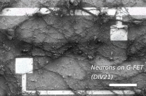
Representative scanning electron micrograph of 21 days old hippocampal neurons interfaced with the G-FETs. The 250 μm-long graphene strip appears darker between the two bright metallic electrodes.
For organic semi-conductors that are used for solar cells we study the different step through which the incident light energy is transformed first in chemical (excitonic) energy and then in electrical energy. The fundamental mechanism that are in play here occur also in numerous systems in photo-catalysis and photo-synthesis.
First, the photon is absorbed in the donor material and creates a localized exciton that carries the photon energy. This exciton diffuses up to the interface between donor and acceptor materials. The mechanism of electron-hole separation that occurs at the interface constitutes a key step for the transformation of energy and also for the solar cell efficiency. The interface is a complex zone and the electron-hole separation depends on the molecule on both sides, on the local electrical potential and on the coupling between charges and vibration modes.
In the first step of photosynthesis (light part), there is also a formation of exciton by photon absorption and then a charge separation between the electron and the hole that are bound in the exciton. Yet the separated charges do not create an electrical current as in a solar cell but rather produce series of redox reactions.
.

(1) An exciton is created on the donor side (left) by photon absorption; (2) It diffuses up to the donor-acceptor interface; (3) It splits into an electron in the acceptor side (right) and a hole in the donor side (left).
We develop various magnetic field sources for applications in life sciences. These field sources are of two kinds: (i) micro-magnets in the 10-100 µm range, which produce magnetic field gradients of up to 106 T/m and therefore considerable forces at the cellular scale and (ii) pulsed magnetic field generators that produce magnetic fields of up to 10 T over 10 µs (Linksium projet pumag).
In collaboration with biologists, we use these field sources to study the influence of magnetic fields on cells and cellular processes (coll. Czech Academy of Sciences, Prague; PLoS ONE, 2013, 8, e70416) and to mechanically manipulate a range of biological entities (cells, embryos, DNA…). This manipulation can serve different purposes:
• Magnetic cell trapping by micro-magnets (coll. Ampere, Lyon; Sensors and Actuators B, 2014, 195, 581)
• Applying physiological stress on embryos through the interaction between micro-magnets and internalised magnetic nanoparticles (coll. Institut Curie, Paris; Nature Comm., 2017, 8,13883)
• Deformation of individual cells by a magneto-active substrate containing micro-magnets (coll. LIPhy, Grenoble; Rep., 2018, 8, 1464)
• Magnetic micro-robots for cell displacement (coll. Liphy and G2ELab, Grenoble).

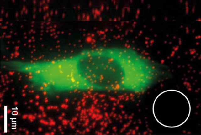
(Left) trapping of magnetic cells by NdFeB micro-magnets observed in fluorescence; (middle) magneto-active substrate for local mechanical stimulation of single cells; (right) magnetic micro-gripper for cell manipulation.
The development of optical tweezers has been mostly driven by their high potential for life sciences. It has led to significant improvements in the manipulation and characterization of cells, proteins and DNA.
At Institut Néel, we have developped different kind of fibered optical tweezers. Compared to the optical tweezers integrated into a microscope, they are particularly relevant in complex environment or large volumes. We recently developed micro-structured fibered optical tweezers that allow efficient trapping with either two face-to-face fibers, or a single fiber. With the latter, we managed to trap yeast, bacteria and living algae.
Since it has been proved that genetics does not drive the full biological development at cellular level but that mechanical, physical and chemical environments have a great influence on cell development, it becomes highly relevant to study these environmental impacts. For this purpose, we use AFM (Atomic Force Microcopy) to measure semi-quantitative force maps on individual cells and we combine them with fluorescent maps that provide 3D architectural information.
Then we merge these set of experimental characterizations in a virtual reality (VR) engine that is equipped with a haptic system to develop an interactive VR station to explore dynamically the data. This multi-sensorial station is part of “enhancing data” approach improving the exploration of complex and multiple data.
These studies are developed in collaboration with biologists from IAB (Institut pour l’Avancée des Biosciences, Grenoble), computing and robotics scientists of ICA (Ingénierie et Créaction Artistique- Grenoble) and ENISE (Ecole Nationale d’Ingénieurs de Saint-Etienne) and with the technological support of the Nanoworld platform of CIME Nanotech.
We have developed a home-made setup dedicated to optical correlation spectroscopies experiments for in situ characterization of metallic or fluorescent colloidal solutions. The goal of such a tool is to measure the diffusion coefficient of the nanoparticles in solution to determine their size and/or their aggregation state which are key parameters for the physical and chemical properties of the colloidal solutions. DLS (Dynamic Light Scattering) experiments are performed by analyzing the thermal fluctuations of the nanoparticles Rayleigh scattering which permits to characterize the solution as a whole. FCS (Fluorescent Correlation Spectroscopy) and SERS-CS (Surface Enhanced Raman Scattering Correlation Spectroscopy) can also be realized by analyzing the thermal fluctuations of the fluorescence of the nanoparticles or the SERS signal of molecules adsorbed on metallic nanoparticles (ACS Omega 2019, 4 p 2283). Such approaches confer a chemical selectivity that enables to target the nano-objet to be studied which is a significant issue in biology.
Nanomaterials engineering is used to produce original nanotracers: nanocrystals made of organic fluorophores with high two-photon absorption cross section and emitting in the red/infrared (1st biological window) are being investigated for in vivo bio-imaging. They are produced using an original one-step spray-drying process as silica-coated nanocrystals which can be easily functionalized (New J Chem 2018 p15353). Furthermore, fluorescent nanocrystal colloids are also being produced in solution using a home-made sonocrystallization setup, and exhibit very high brightness in vitro. (coll. C Andraud, ENS Lyon; M Gary-Bobo, IBMM Montpellier and O Pascual, INSERM Lyon).
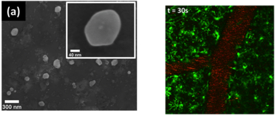
(Left) Organic nanocrystals after etching of the silicate matrix; (Right) Two-photon fluorescence image of the vasculature of a mice brain using fluorescent organic@silicate nanoparticles (red). The mice expresses GFP in the microglia (green).
We are also strongly involved in the preparation of inorganic nanocrystals for bio-imaging using photoluminescence in the near-infrared and nanothermometry applications.
We developed, through a microwave-assisted solvothermal route, iodate nanocrystals (such as α-La(IO3)3 doped with Yb3+ and Er3+), which exhibit both efficient NLO signal and photoluminescence under a single excitation wavelength (Coll. SYMME Annecy), paving the way to new multifunctional nanoprobes for biophotonics (Inorg. Chem. 2019, 58 p1647 and Cryst. Eng. Comm. 2020, 22 p2517).
Meanwhile, we synthesize, still using solvothermal process, oxide garnet-type nanocrystals doped with Nd3+ (or co-doped with different ions), presenting photoluminescence in the near-infrared range, i.e. in the biological transparency windows for in vivo bio-imaging (coll. Univ. Madrid)
Both iodate and oxide nanocrystals may also find applications for thermal imaging.
Yb,Er-doped nano-iodates are currently studied for in vitro temperature measurements in neurons (coll. Cécile Delacour, Valérie Reita), with the goal of monitoring weak temperature changes (0.2K) at the single cell level. These nanocrystals evidence a thermal sensitivity of 1.2 %.K-1 in the green range, when monitoring the (4S3/2, 2H11/2) → 4I15/2 levels of Er3+, even in the presence of neurons.
Nd3+-doped garnet-type nanocrystals (YAG and Gd3Sc2Al3O12) have a high potential as nanothermometers in the physiological temperature range (20-50°C), through thermally coupled sublevels of 4F3/2→4I9/2 electronic transition of Nd3+. Indeed, we registered a higher thermal sensitivity (0.20 % K-1) compared to that of Nd3+ doped YAG nanoparticles, due to the difference in crystal field of the host matrices (PCCP 2019 p11132; coll. INRS-Montreal). Other oxide matrices, or borates, are currently investigated in this nanothermometry field in collaboration with Prof. Lauro Maia (Univ. Goias, Brazil).
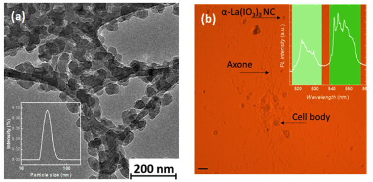
(a) Low temperature TEM image of Yb,Er-doped α-La(IO3)3 nanocrystals. In the insert, the DLS size distribution in ethylene glycol. (b) Optical image of neurons with α-La(IO3)3 nanocrystals (Scale bar: 2 µm). In the set, emission spectrum of Yb,Er-doped α-La(IO3)3 nanocrystals.
Finally, mesoporous SiO2 and core-shell Au@SiO2 nanoparticles are being developed for the encapsulation and delivery of proteins. Au@SiO2 nanoparticles should serve as delivery tools for DNA-repairing enzymes for early cancer treatment (IDEX funding, Coll. S Aldabe Bilmes, Univ Buenos Aires and Y Roupioz, SyMMES Grenoble), while large-pore mesoporous silica nanoparticles functionalized with DNA aptamers are being developed for the delivery of antimicrobial peptides to fight multi-resistant bacteria (coll. F Oukacine DPM Grenoble).
We have assessed the potential of graphene-on-polymer films for biomedical and bioelectrical applications. It has led to the realization of several “smart-bandage”prototypes with promising perspectives for wound healing. The marked interest of the medical community has motivated the creation of “Grapheal”, a spin-off company that has been hosted by Institut Néel, that is aiming at developing a range of medical devices combining embedded electronics with graphene technology in order to reach the market of advanced woundcare.
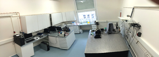
Biotechnological platforms for cells observations, manipulation and culture with epifluorescent microscopes, fast imaging and Patch Clamp station for electrophysiology, microfluidic devices, and culture room (class P2).

In Transmission Electron Microscopy, Institut Néel occasionally collaborates with Grenoble biology laboratories: cryogenic experiments carried out at the IBS on samples from the Néel Institute, microanalysis experiments carried out at Institut Néel on biological samples from ISTerre. For several years, the microscopists of Institut Néel have been developing observation techniques in low-dose mode, intended for fragile samples.
Field emission scanning electron microscopy (FESEM) allows to observe micro and nano objects with a spatial resolution about one or few nanometers depending of the nature of them. By using a very low voltage irradiation damage can be limited and it is possible to observe cells or strands of woven DNA.
Sometimes, it is necessary to make a conductive thin film (around 1 nm) to limit charges effects by Physical Vapour Deposition (sputtering). The FESEM of the Institut Néel works in a range of 100 V to 30 kV (spatial resolution: 1 nm at 15 kV and 1.7 nm at 1 kV. Thin films can be done from 1nm or more with Gold, Platinum, Aluminum, Carbon or ITO (In2-xSnxO3).
Since 2015, Institut Néel is a member of the LANEF, which is a Laboratoire d’Excellence from Grenoble supporting projects around nanosciences and energy. In particular, one branch of the LANEF is dedicated to Nanosensors and nanomaterials for healthcare and biology. Beyond financial support for PhDs, some thematic workshop are also organized in the framework of LANEF to favour exchange and networking between the member labs.
The SATT Linksium has accompanied several biology-related projects from Institut Néel through maturation, incubation and eventually creation of a company. Among them, Grapheal (graphene-based smart bandages), Pumag (pulsed magnetic fields), and Pumpit! (magnetic micropumps).
