Technological hub
Collaborating Research Group at ESRF/ILL facilities
Exploitation et développement des instruments des CRG dédiés à la science des matériaux et hébergés dans les installations européennes ILL et ESRF
CRG et grands instruments:Goals
The CRG pole, with researchers and engineers from Institut NÉEL, CEA/IRIG, OSUG and CNRS-SIMaP, is in charge of the operation and development of CRG instruments hosted at ILL and ESRF. They drive and supervise experiments for national and international scientific teams. Beamtime is allocated by review committees.
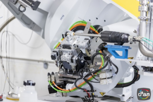
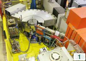
D1B at ILL, is a high flux neutron powder diffractometer dedicated to materials science with emphasis on magnetism and in situ / operando experiments.
The 5 French CRG beamlines at ESRF (f-crg.fr) cover 4 main domains : Earth and Environment, Biology and Health, Materials and Energy, Communication and Information, with in situ/operando characterization tools (HR-XRD, EXAFS, SAXS/WAXS…).
 |
 |
Introduction
D1B is a French-Spanish CRG (Collaboratin Research Group) instrument run by CNRS and CSIC at the Institut Laue-Langevin.
D1B Diffractometer
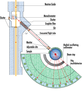 D1B is a high intensity powder diffractometer with an 128° PSD.
It has always been in very high demand for real time experiments, and for very small samples because of its high efficiency position sensitive detector (PSD).
A great number of experiments performed on D1B concern the determination of magnetic structures. At small angles where the magnetic peaks are expected, a high spatial resolution can be achieved, the FWHM reaches 0.2° (for a sample with = 8 mm).
The characteristics of the instrument and of its applications are presented on the web site of ILL : CRG-D1B
Submit a proposal
Since the birth of the Fédération Française de Diffusion Neutronique (2FDN) the call for proposals at D1B are managed together with all the LLB instruments and the others CRG at the ILL.
There are two call for proposal per year, one in spring and the other in autumn. The proposal must by submitted via the Phoenix website. You can contact the CRG team for preparing your proposal at the address crg-d1b@grenoble.cnrs.fr
Contacts :
– inquiries concerning the feasibility of an experiment, the instrument performances and the sample environments : Vivian NASSIF
– questions about the studies of magnetic structures, nuclear/magnetic phase transitions : Claire COLIN
– questions about in-situ experiments: Lætitia LAVERSENNE
Introduction
The French CRG (Collaborating Research Group) lines at the ESRF received in 1994 from the CNRS and the CEA the missions of strengthening and facilitating the use of synchrotron techniques through, on the one hand, the support and training of scientists from French laboratories, and secondly the development of advanced instrumentation and analysis tools.
These five beamlines, D2AM (anomalous diffraction / scattering for materials science), IF (diffraction / scattering at surfaces and interfaces), FIP (crystallography of biological macromolecules), FAME (absorption spectroscopy for earth sciences and environment) and FAME-UHD (idem, but on ultra-diluted metallic trace elements) are currently installed on ports BM02, BM32, BM07, BM30 and BM16 of the ESRF.
For more details go to the f-crg website.
D2AM – BM02
Multi-wavelength Anomalous Diffraction and Scattering
D2AM is a beamline dedicated to in situ or operando materials science: reciprocal space systematic exploration, measurement of weak diffraction or scattering signals, at large and small angles, use of the anomalous effect.
One can conduct fundamental or applied research on all aspects of the materials complexity: atomic structure, defects, chemical order/disorder, microstructures, scales hierarchy, heterogeneity.
The new Kappa diffractometer and scattering bench can analyse samples in various environments: low and high temperatures, electro-chemistry, liquid process growth, tensile machine…
The new X-Ray optics delivers a stable monochromatic flux with the following characteristics:
- 80 x 150 μm2
- de 6 à 40 keV
- flux of 1011 photons/s on the sample.
The use of 2D hybrid pixel XPAD detectors allows extremely fast acquisitions.
DOMAINES D’ÉTUDE
- Nano-structured thin films and nano-materials
Deformation fields, chemical composition: nano-wires, magnetic alloys, micro-electronic materials, IT technologies. - Multi-scale and heterogeneous materials
Distortion, segregation, microstructure, defects, composition: metallic alloys, under temperature and external constraints, ceramics, cultural heritage, composite materials. - Polymers and solutions
Morphology, organisation, ageing and constraints, cavitation: doped polymers, nanoparticules, life interfacing poly-electrolytes.
TECHNIQUES
- Diffraction, Scattering, in grazing incidence
- Multi-wavelength diffraction spectroscopy
- Anomalous scattering at small and large angles
IF – BM32
Surfaces et Interfaces
The IF (InterFaces) beamline is dedicated to structural and applied studies in nanoscience and technologies using the synchrotron radiation.
Materials can be studies ex situ or in situ (under high ultra vacuum, or controlled atmosphere, in air) during their elaboration & working processes or under various loadings (e.g.: thermal, mechanical, electrical).
3 INSTRUMENTS
- Ultra-vacuum in situ nanostructures studies instrument
Atomic structure, morphology and composition.
Nano-structures : islands, particles, nanowires, graphene, 2D materials.
Surfaces-interfaces : alloys, heterostructures, catalyse.
Growth: Molecular-beam epitaxy, chemical vapor deposition. - The multi-techniques goniometer
Interfaces solid/solid : adhesion, implantation, molecular sticking.
Interfaces liquide/solide : microfluidics, surface alteration, Li-ion battery.
Thin layers, properties-structure relationship in functional materials. - The multi-techniques goniometer
Local orientation and deformation map with submicrometric resolution.
3D integration in micro-electronics.
Control and reliability of materials for energy (nuclear and renewable).
Biology-health materials.
Mechanical tests on micro- and nano-objects.
TECHNIQUES
- Diffraction, Scattering, in grazing incidence
- X reflectivity
- Laue Microdiffraction
FAME – BM30
Absorption spectroscopy, Earth Science and environment
FAME is dedicated to x-ray absorption spectroscopy. Thanks to the combination of photon flux optimization, stability and fluorescence detection, the installation offers the possibility to probe chemical elements with concentrations ranging from 50ppm to 100%, in various conditions: in situ (high pressure and high temperature), operando (cell for electrochemistry or catalysis), at low temperature (for samples sensitive to beam damage or for phase transition studies).
TECHNIQUES
- Ex situ and in situ chemical speciation of trace elements by x-ray absorption spectroscopy
- Modes : transmission & fluorescence
- Techniques : XANES & EXAFS
FAME-UHD – BM16
SHigh resolution / high sensitivity absorption spectroscopy
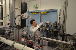
The multi-crystal analyser spectrometer on FAME-UHD
FAME-UHD is dedicated to x-ray absorption spectroscopy in ultra-diluted conditions, for the study of trace elements in samples with environmental, chemical or biological interest. EXAFS analyses can be carried out at concentrations as low as 10ppm and XANES analyses down to less than 1ppm. To do so, a spectrometer constituted of 14 bent crystal analyzers has been built. This type of high resolution detector brings improvements and progresses to the research carried out on the installation through two directions : i) study more and more complex samples and ii) improve and enrich the information obtained about the electronic structure of the probed elements.
Thus, several new scientific opportunities are offered to the user:
- it becomes possible to carry out EXAFS fluorescence measurements on diluted elements in matrices with main constituants which are excited by the incident beam, with a lowered detection limit,
- the resolution of the absorption edge becomes much more precise (high resolution XANES),
- the fine study of the x-ray emission spectrum of the element of interest as a fonction of the energy of the incident photons becomes possible as well (RIXS measurements, Resonnant Inelastic X-ray Scattering).
TECHNIQUES
- XAS, EXAFS, XANES, XES, HERFD-XAS
FIP2 – BM07
Biological macromolecular crystallography
This beamline is specially dedicated to the crystallography of biological macromolecules over a wide range of wavelengths, with the possibility of using the anomalous signal.
It also allows you to work at room temperature.
Thanks to this tool, it is possible to reveal the three-dimensional structure on the atomic scale of biological macro-molecules in order to answer fundamental questions, but also to address therapeutic or industrial issues.
TECHNIQUES
- Macromolecular crystallography
- Multi-wavelength anomalous scattering
- “In situ” diffraction (using drop plate)
- “On-line” spectroscopy